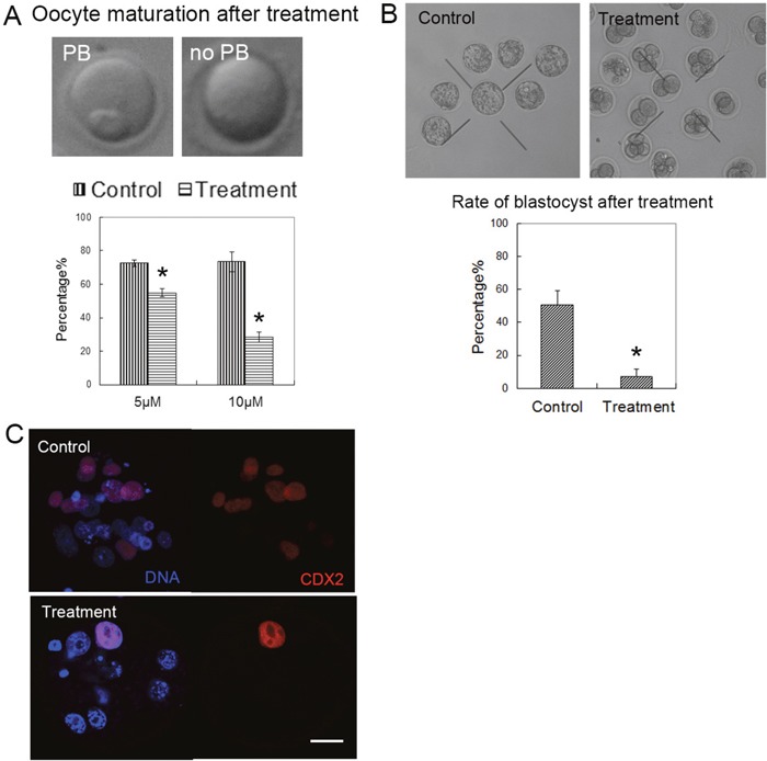
Figure 1. Citrinin toxin exposure affected oocyte maturation and early embryo development.

(A) The oocytes were cultured with 5 μM, 10 μM citrinin toxin for 12 h. Most oocytes developed to the MII stage in the control group, however, most of oocytes failed to reach to the MII stage. (B) The blastocyst embryo rate was significantly reduced with citrinin toxin treatment. *, significantly different (p < 0.01). (C) The expression of CDX2 in the embryos. In the control embryo, CDX2 expressed in the TE cells; while in the treated embryo, there are barely CDX2 signal. CDX2, red; DNA, blue. Bar = 20 μm.
