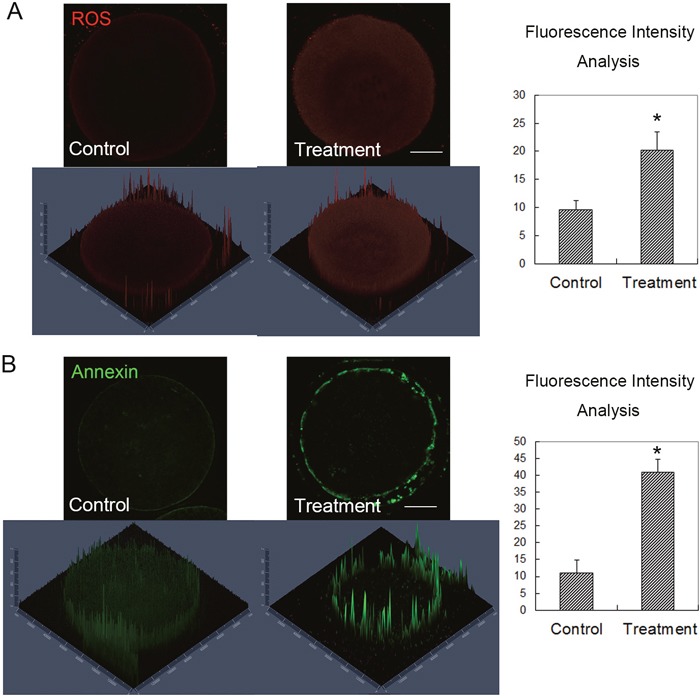
Figure 4. Citrinin toxin exposure induced oxidative stress and early apoptosis in mouse oocytes.

(A) After citrinin toxin treatment, the ROS level was significantly increased in the treated oocytes, while the average ROS fluorescence intensity analysis showed that ROS signal increased significantly in the treated mouse oocytes. *, significantly different (p < 0.05), Bar = 20 μm. (B) After citrinin toxin treatment, the Annexin signal, which presented the early apoptosis level was significantly increased in the citrinin treated oocytes, while the average Annexin fluorescence intensity analysis showed that Annexin signal increased significantly in the treated mouse oocytes. *, significantly different (p < 0.05), Bar = 20 μm.
