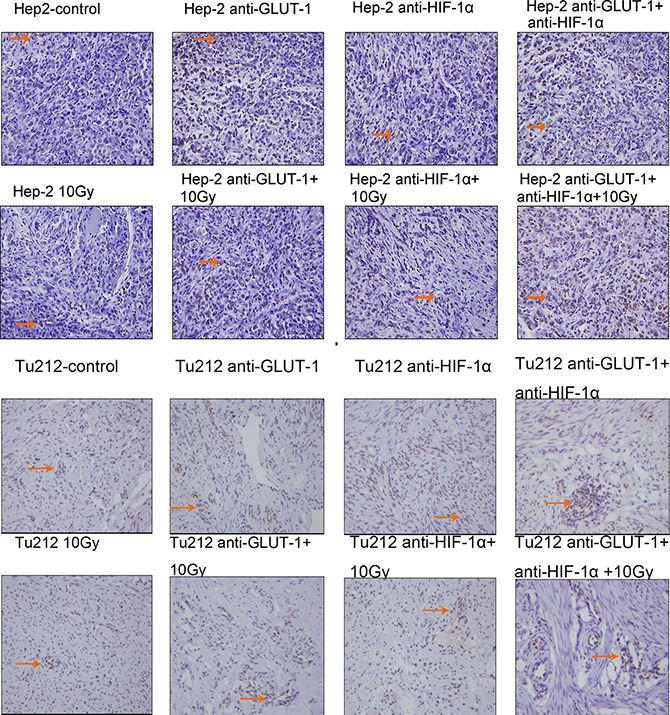
Figure 3. TUNEL positive staining showed that the nucleus was brown or brown-yellow, that is, apoptotic cells.

The arrows in the figure refer to apoptotic cells of Hep-2 and Tu212 xenografts 8 days after treatment. Apoptosis index were observed under an optical microscope (magnification, ×400).
