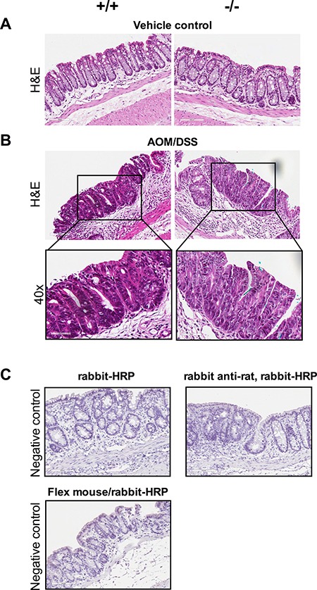
Figure 2. Dysplastic lesions/ACFs developed in AOM/DSS mice independent of Cysltr1 status.

The colons of vehicle control and AOM/DSS mice with different Cysltr1 genotype were processed for immunostaining. (A) Representative images (20×) of hematoxylin and eosin staining of vehicle control colons with no observed polyps. (B) Representative images (20× and 40×) of AOM/DSS female mice with different Cysltr1 genotypes with dysplastic lesions/ACFs stained for hematoxylin and eosin (n = 3–6 per genotype). (C) Negative controls (IgG controls) for all the secondary antibodies used, rabbit-horseradish peroxidase (HRP), rabbit anti-rat with rabbit-HRP, and combined Flex mouse/rabbit-HRP.
