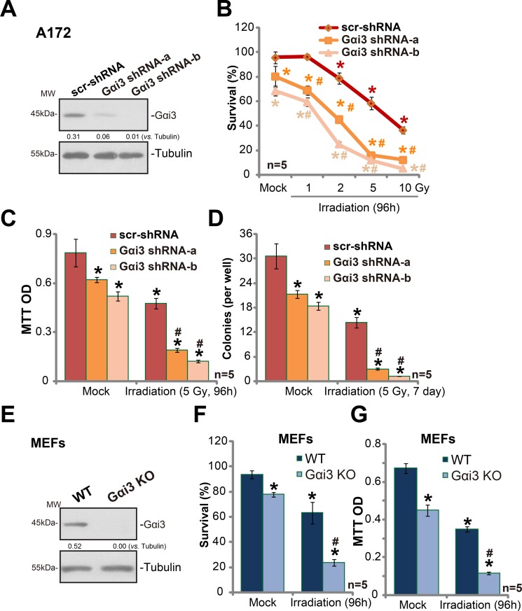
Figure 1. Silencing Gαi3 sensitizes irradiation-induced glioma cell death.
Western blotting tested expression of listed proteins in stable A172 cells with Gαi3 shRNA (“−a/−b”) or scramble control shRNA (“scr-shRNA”) (A); A172 cells were also subjected to irradiation (at indicated intensity) and cultured for indicated time, listed assays were performed to test cell survival/death (B–D). Expression of listed proteins in wild-type (WT) and Gαi3 knockout (KO) MEFs was shown (E); MEFs were irradiated (5 Gy) and cultured for additional 96 hours. Afterwards, MEFs were subjected to trypan blue staining assay (F) and MTT assay (G). For all the assays, the exact same number of viable cells of different genetic background was initially plated into each well (Same for all Figures). Same set of lysate samples were run in sister gels (A and E). “Mock” stands for un-irradiated cells (Same for all Figures). “n = 5” means five replicate wells (Same for all Figures). Bars stand for mean ± SD (Same for all Figures). *p < 0.05 vs. “Mock” of “scr-shRNA” A172 cells or WT MEFs. #p < 0.05 vs. “Irradiation” of “scr-shRNA” A172 cells (B–D) or WT MEFs (F and G). Experiments in this figure were repeated three times, with similar results obtained.

