
Figure 5. The expression of apoptosis-related genes through RUNX3 downstream by Pa-PDT.
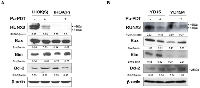
(A-B) Cells were treated with Pa-PDT (0.3 μM Pa and 0.5 J/cm2). After 4 h, the cells were collected and the protein expression of RUNX3, Bax, Bim, and Bcl-2 were measured by western blotting. Cropped gels retain suitable bands for each of primers and antibodies. The relative intensity of protein expression was measured by Image J software.