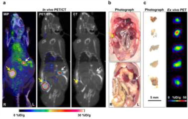
Fig. 5.

Radiological-surgical correlation of SKOV3 orthotopic ovarian cancer model. (A) In PET/CT imaging, a primary tumor with strong uptake was found in the right ovary area (yellow arrow), and there were many focal uptakes in the peritoneal space (white arrow). (B) Surgical exploration was done in the same animal after terminal PET/CT imaging. The primary tumor mass was found in the right ovary (yellow arrow), and there was white nodular tissue (white arrow) at the location of focal uptake on the PET/CT imaging, suggesting peritoneal implants. (C) Ex vivo PET imaging of excised peritoneal implants was performed. The small peritoneal implants showed high PET uptake values.
