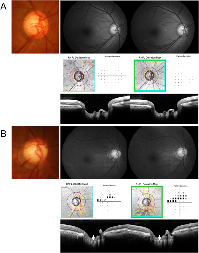
Figure 2.

Representative cases. (A) A 56-year-old female with normal-tension glaucoma. A inferotemporal localized retinal nerve fiber layer (RNFL) defect with disc hemorrhage (DH) is shown in the right eye. Enhanced depth imaging (EDI) of the optic nerve head shows no focal lamina cribrosa (LC) defects. During five years of follow-up, there was no evidence of glaucoma progression in this case. (B) A 69-year-old male with normal-tension glaucoma. Diffuse RNFL defect with inferotemporal DH is shown in the right eye. Enhanced depth imaging (EDI) of the optic nerve head shows a focal lamina cribrosa (LC) defect at the site of DH. During four years of follow-up, there were progression in both the RNFL and the visual field.
