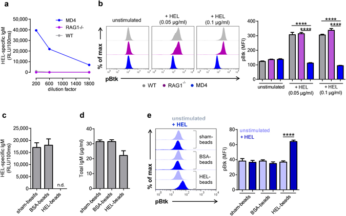
Figure 4.

Antigen-specific secreted IgM limit self-antigen induced B cell receptor signaling. (a) Quantification of hen egg-white lyoszyme (HEL) specific IgM in the plasma of MD4, RAG1−/− and wild-type (WT) mice by ELISA. (b) Representative flow cytometry plots and bar graphs represent the mean fluorescence intensity (MFI) for pBtk in purified B-2 (B220+ CD43−) cells from MD4+/− mice stimulated with HEL for 3 minutes in presence of either WT (grey), RAG1−/− (purple) or MD4 plasma (blue). ELISA quantification of (c) HEL-specific and (d) total IgM in MD4 plasma treated with either unconjugated or BSA- or HEL-conjugated microspheres. (e) Representative flow cytometry plots and bar graphs show the MFI for pBtk in purified B-2 cells from MD4+/− mice stimulated with HEL for 3 minutes in the presence of plasma from MD4 mice that has been treated with either unconjugated or BSA- or HEL-conjugated microspheres. All results show mean ± SEM, ****P < 0.0001 (unpaired t test or One-Way Anova followed by Tukey’s test). n.d.: not detechtable.
