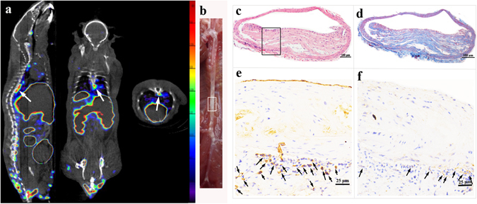
Figure 8.

In-vivo SPECT/CT imaging and pathological results. (a) In vivo micro-SPECT/CT images of 99mTc-MAG3-bevacizumab (sagittal, coronal, and transverse views, left to right) in an ApoE−/− mouse. (b) The gross morphology of aortic plaque. (c) IHC results of slices from intense radioactivity accumulated plaque area in aortic arch, CD31 immunohistochemical image (×100), higher magnification (×400), VEGF immunohistochemical image (×100) and higher magnification (×400) displayed from left to right in turn. (d) IHC results of slices from intense radioactivity accumulated plaque area in thoracic aorta, CD31 immunohistochemical image (×100), higher magnification (×400), VEGF immunohistochemical image (×100) and higher magnification (×400) displayed from left to right in turn.
