Figure 1.

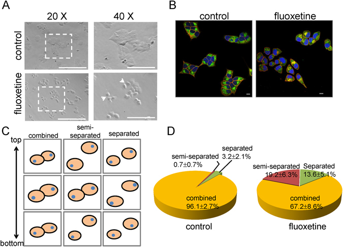
Fluoxetine alters cell morphology, and reduces cell-cell adhesion. (A) After 3-hour fluoxetine (30 μM) treatment, MIN6 cells were observed under an inverted fluorescence microscope (Evos). The white arrows indicate reduction of cell-cell adhesion. Scale bar, 100 μm. The representative images were from at least three independent experiments. (B) After 3-hour incubation with or without fluoxetine (30 μM), MIN6 cells were fixed and then immuno-stained with Alexa 488 (green) for E-cadherin, Alexa 594 (red) for β-catenin and Hoechst 33258 (blue) for nucleus. The images were captured by using confocal microscope (Olympus, MPE). Scale bar, 10 μm. The representative images were from at least three independent experiments. (C) Schematic diagram defines three characteristics of cell contact. Cells were categorized by how close they contact to each other at different z-sections. Cell junction was fully continuous from top to bottom (“combined” cells), partially lost at the top and bottom (“semi-separated” cells) or totally lost from top to bottom (“separated” cells). (D) Quantitative analysis for the percentage of each contact type of MIN6 cells treated with or without fluoxetine. Each value represents mean ± SEM of at least 600 individual cells.
