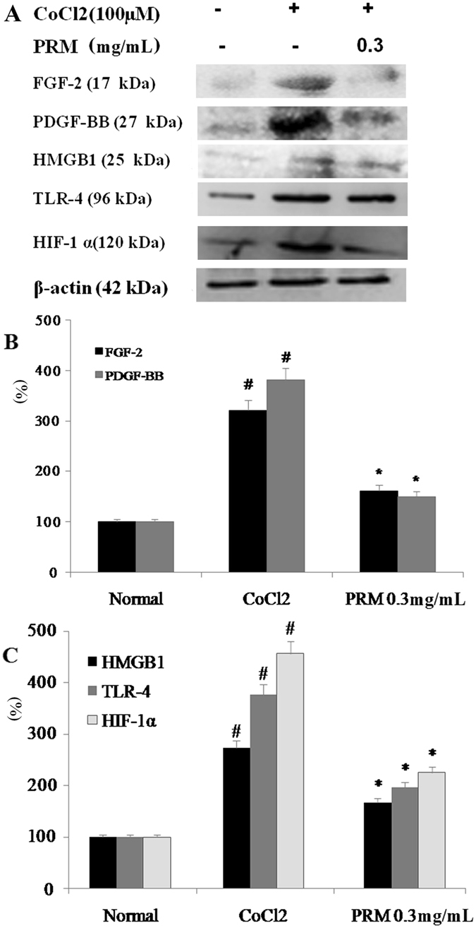
Figure 5.

Effects of PRM on HIF-1α, TLR-4, PDGF-BB and FGF-2 expression in CoCl2 stimulated HLF-1. (A) Representative light microscopic appearance of normal HLF-1 cells (A1), CoCl2-treated cells (A2), and cells treated with CoCl2 +0.3 mg/mL PRM (A3). (B and C), HLF-1 cells were incubated with CoCl2 (100 μM) for 48 h. HMGB1, HIF-1α, TLR-4, PDGF-BB and FGF-2 expression was analyzed by western blot. The results are reported as percent increase over normal. Data are reported as the mean ± S.D., n = 5. #P < 0.01 vs. the normal group; *P < 0.05, **P < 0.01 vs. the CoCl2 stimulated group. Significance was determined by one-way ANOVA followed by Dunnett’s test. The full-length blots of Fig. 5A are presented in Supplementary information 2.3.
