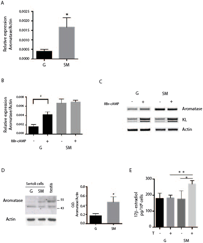
Figure 4.

SM increases P450-aromatase expression in Sertoli cells. Sertoli cells were cultured for 48 h at G or in RCCS. (A) Real time PCR of P450-aromatase showing a strong increase of mRNA level under microgravity condition. (B) Real time-PCR for P450-aromatase in Sertoli cells treated or not with 1 mM 8-Br-cAMP. cAMP inducibility is lost under SM. (C) Semiquantitative RT-PCR of P450-aromatase and KL in Sertoli cells treated or not for 48 h with 1 mM 8-Br-cAMP. P450-aromatase expression is stimulated by 8-Br-cAMP at G but not under SM, while KL expression is stimulated by 8-Br-cAMP in both culture conditions. (D) Western blot analysis of P450-aromatase in Sertoli cells after 48 h of culture at G or under SM. Histogram on the right reports the mean of densitometric analysis of four different experiments. Values were normalized by reference to values for actin. (E) Chemiluminescence immunoassay for estrogen level in the culture medium of Sertoli cells. Sertoli cells were cultured in the presence or absence of 50 nM testosterone at G or in SM for 48 h. SM causes an increase of estrogen in the medium when cells are treated with testosterone. At least three different experiments were performed. Bars represent s.d. Asterisks indicate: *P < 0.05 and **P < 0.01.
