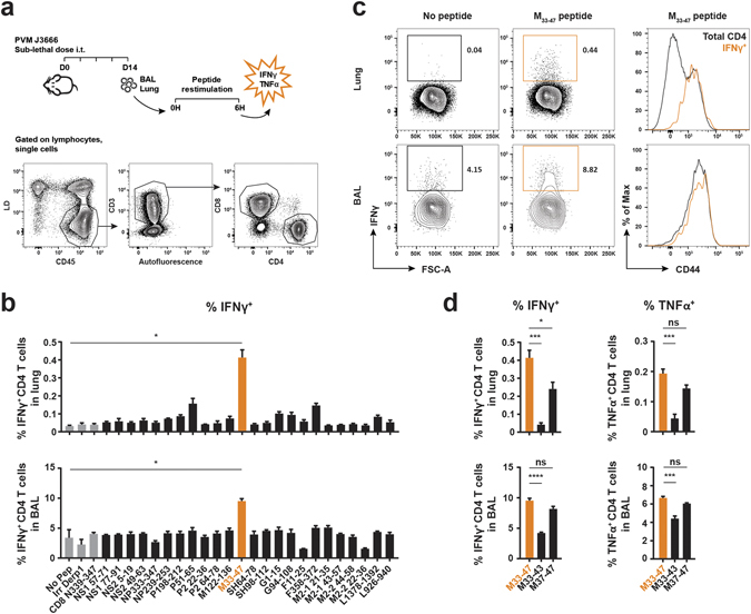
Figure 2.

Identification of MHCII-restricted PVM epitopes recognized by CD4 T cells from PVM-infected mice. 8 week old C57BL/6 females were infected i.t. with a sub-lethal dose of PVM strain J3666 and sacrificed 14 days later. BAL and lung single-cell suspensions were restimulated for 6 h in the presence of Golgistop and PVM-specific peptides. T cells were evaluated for cytokine production by intracellular staining and flow cytometry analysis. (a) Schematic overview of the experimental setup and gating strategy to identify CD4 and CD8 T cell populations in BAL and Lung. (b–d) IFNγ or TNFα production by CD4 T cells, isolated from lung or BAL, after incubation with each of the predicted MHCII-restricted PVM peptides enlisted in Table 1. As negative controls, cells were incubated without peptide, with an irrelevant Derp1 CD4 peptide27, or with a MHCI-restricted PVM N339–347 peptide20 and are shown in gray. (b) IFNγ production by CD4 T cells in BAL and Lung. Data are depicted as frequency of IFNγ-producing cells among total CD4 T cells. (c) Left, representative FACS plots (gated on CD4+ cells) show the percentage of IFNγ+ CD4 T cells in response to restimulation with or without M33–47 peptide. Right, Histogram overlays depict CD44 expression levels of total CD4+ and gated IFNγ+ CD4+ populations (marked orange in left panel) following restimulation with M33–47 peptide. Data are normalized to and depicted as the percentage of the maximum count (% of max on the Y axis). (d) IFNγ and TNFα production by CD4 T cells following restimulation with M33–47, M33–43, or M37–47. Results are shown as mean ± SEM from three biological replicates. For each biological replicate 10 mice were pooled to obtain sufficient cell numbers for epitope screening. Data are representative of two independent experiments. For statistics, conditions restimulated with peptide were compared to the no-peptide control (b, Student’s t test with Welch correction) or the M33–47 peptide (d, ANOVA for multiple comparisons) as indicated. BAL, bronchoalveolar lavage; IFNγ, interferon gamma; TNFα, tumor necrosis factor alpha; MHCI/II, major histocompatibility complex class I or II; Derp1, Dermatophagoides pteronyssinus peptidase 1; Irr, irrelevant; ns, not significant.
