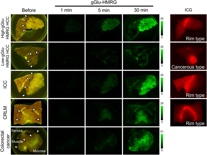
Figure 1.

Fluorescence imaging of HCC, ICC, and CRLM using gGlu-HMRG. Fluorescence images of representative resected tumor samples acquired before and at 1, 5, and 30 min after spraying with gGlu-HMRG. Images were obtained with the blue filter setting (excitation 445–490 nm, emission 515 nm, long pass). The High-gGlu-HMRG HCC, ICC, and CRLM were identified from 5 min after topical administration of gGlu-HMRG, while Low-gGlu-HMRG HCC remained unidentifiable even at 30 min after the imaging. Arrows indicate boundaries of the tumors by macroscopic gross examination. Right-most column shows fluorescence images of the same tumor samples acquired by near-infrared imaging (excitation 710–750 nm, emission 810 nm, long pass). “Cancerous type” indicates a fluorescence signal from the tumor and “rim type” indicates a fluorescence signal from the non-cancerous hepatic parenchyma but not the tumor itself.
