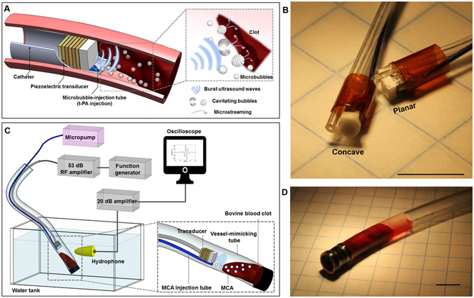
Figure 1.

Microbubble-mediated intravascular sonothrombolysis system. (A) Principle of microbubble-mediated intravascular sonothrombolysis by using a catheter-tipped forward-looking transducer; low-frequency ultrasound waves generate stable and inertial cavitation of locally injected microbubbles near the target blood clot. (B) Customized 620 kHz stacked piezoelectric transducers integrated with a microbubble-injection tube and polyimide housing: concave- and planar-aperture (lateral dimension <1.5 mm) prototypes (scale bar = 5 mm). (C) In vitro experiment setup; a tygon tube (ID: 4 mm, OD: 5.6 mm) was used as a vessel-mimicking structure. The hydrophone was used to detect the microbubble response. (D) Bovine blood clot sample (200 mg ± 10%) located in the tygon tube with 50 μl saline water (scale bar = 5 mm). The figures (A) and (C) were drawn through the combined use of Solidworks Education Edition, Microsoft PowerPoint, and Adobe Illustrator CC. The figures (B) and (D) were captured using a digital SLR camera (Canon EOS Rebel T3) with 50 mm lens (Canon, f/2.5 Compact Macro Lens), cropped and aligned using Microsoft PowerPoint and Adobe Illustrator CC.
