
Figure 3.
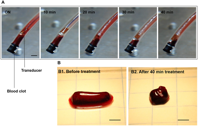
In vitro clot lysis by microbubble-mediated intravascular ultrasound. (A) Captured images during 40 min treatment by using the planar-aperture prototype (input conditions of 620 kHz, 80 Vpp, 10% duty cycle with 100 μl/min injection of 508 bubbles/ml). A smaller (120 mg) clot was used for monitoring the volume reduction (scale bar = 3 mm). Due to the high bubble concentration (508 bubbles/ml) we observed that the undestroyed bubbles were attached to the transducer and the inner surface of the tygon tube. (B) A 120 mg clot was reduced to 50 mg after the 40 min treatment (scale bar = 3 mm).