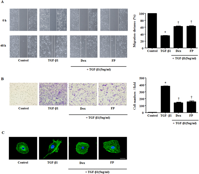
Figure 2.

Glucocorticoids mediate migratory and invasive properties stimulated by TGF-β1 in A549 cells. Cells were pretreated with or without dexamethasone (Dex, 2.5 μM) and fluticasone propionate (FP, 2.5 μM) for 1 hour and then stimulated with TGF-β1 (5 ng/ml). (A) Wound scratch and (B) Transwell migration assays established migration and invasion by A549 cells. Representative images are shown. In the wound scratching assays, the graphic representation is the percent of migrated cells counted within the areas of healing covering the lines present at baseline. In the Transwell migration assays, invasive cells were counted in five high-power fields (HPFs) for the average cell number migrated per HPF. (C) Visualization of F-actin used phalloidin with DAPI staining. Immunofluorescence was determined by confocal laser scanning microscope. Scale bar = 20 μm. Data are presented as means ± SEM. Results were from at least three independent experiments. *p < 0.05 vs. control; †p < 0.05 vs. TGF-β1.
