Fig. 4.

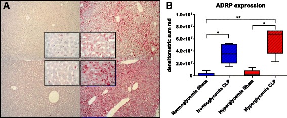
Lipid droplet formation in the liver. a Exemplary pictures for ADRP immunohistochemistry (10×, insets 40×, top left normo sham, top right normo CLP, bottom left hyper sham, bottom right hyper CLP) b ADRP expression, * p ≤ 0.05, ** p ≤ 0.01. All data is presented as median (interquartile range). Kruskal-Wallis analysis of variance on ranks with post hoc Dunn’s test for multiple comparisons
