
Figure 7.
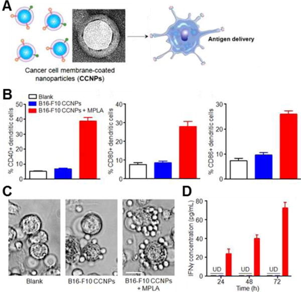
Cancer cell membrane-coated nanoparticles (CCNPs) for anticancer vaccination. (A) Depiction of CCNPs for antigen delivery to dendritic cells. (B) Quantitative flow cytometry data of dendritic cell maturation when incubated for 48 hours with CCNPs coated with membrane from B16-F10 mouse melanoma cancer cells (B16-F10 CCNPs), with or without the adjuvant MPLA. (C) Phase contrast microscopy images of splenocytes derived from pmel-1 transgenic mice when incubated with dendritic cells pulsed with B16-F10 CCNPs, with or without MPLA. Scale bar = 25 μm. (D) IFNγ ELISA of supernatant collected from co-culture at 24, 48, and 72 hours. UD, undetectable by ELISA. Adapted with permission from reference 56. Copyright 2014, American Chemical Society.