Figure 3.
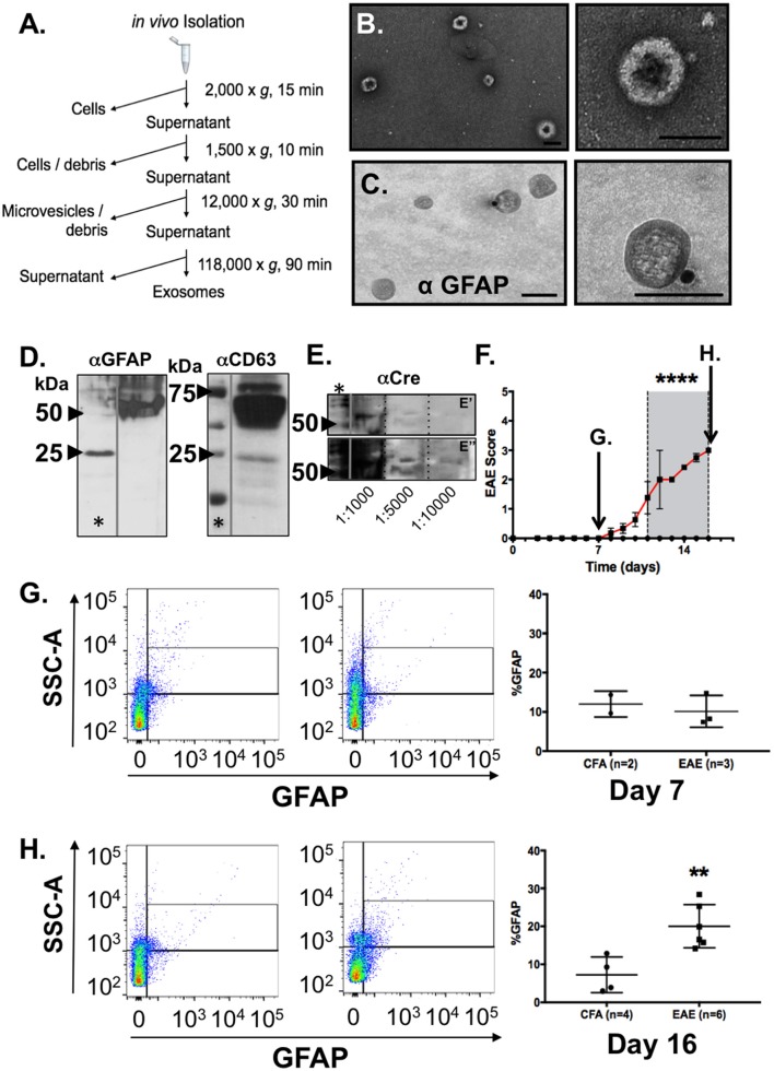
Identification and characterization of astrocyte-derived exosomes in blood plasma from mice and increased detection of GFAP+ exosomes in blood from mice during EAE. (A) Work flow scheme for exosome isolation from blood plasma. (B) Electron micrographs of extracellular vesicles isolated from peripheral blood plasma revealed prototypic “cup-shaped morphology” of the appropriate size class for exosomes. (C) Immunogold electron microscopy using anti-GFAP and gold particle conjugated secondary antisera (15 nm) identified exosomes from blood plasma that were astrocyte-derived. Scale bars: size bar, (A) = 100 nm; (B) = 100 nm. (D) Western blotting analysis of the EM exosome preparations from blood plasma analyzed in (A,B) confirmed detection of exosomal (CD63) and astrocytic markers (GFAP) in the exosome pellet. (E) Validation of astrocytic origins using expression of CRE recombinase protein in blood plasma as measured by western blotting from exosome preparation from GFAP-CRE transgenic mice. Samples were diluted (as indicated) to demonstrate that antibody reactivity was serially diminished which supports the specificity of the antibody binding. (E') represents a shorter exposure of the blot, while (E”) represents a longer exposure time which were needed to visualize CRE-reactive bands in the more diluted samples, respectively. The CRE-reactive bands were of the same molecular weight as observed in the most concentrated sample. (F) Clinical EAE scores of myelin oligodendrocyte glycoprotein (MOG) immunized C57BL/6 mice (n = 6) and control (CFA inoculated; n = 4). Mice were euthanized one week following immunization or time point of peak clinical illness (Day 16). Euthanization of CFA control animals were time-matched for either day 7 (arrow) or day 16 (arrow), respectively, for blood collection and blood plasma exosome isolation. (G) Flow cytometry analysis of blood plasma exosomes from EAE and control (CFA) mice at a pre-clinical disease timepoint (Day 7), and (H) at the time of peak clinical illness (Day 16). GFAP+ exosomes in blood of control mice did not differ in preclinical disease (Day 7) MOG immunized mice, whereas a notable increase in the detection of astrocyte-derived exosomes in blood plasma was observed in mice at the time of peak clinical illness in EAE. Significance is indicated where: (F) ****P < 0.0001, 2-way ANOVA; (H) **P = 0.0031; t-test.

