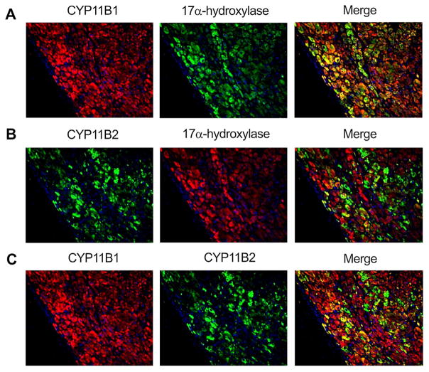
Fig. 3.
Immunofluorescence of CYP11B2, CYP11B1 and CYP17 in the aldosterone-producing adenoma (APA): [A] CYP11B1 and CYP17 were diffusely detected in the tumoral cells of APA. The merged photograph of CYP11B1 and CYP17 is presented on the right. [B] CYP11B2 was detected in patches of tumoral cells in a diffuse manner along-with non-fluorescent CYP11B2 negative cells in-between. CYP17 showed the same pattern described in [A]. The merged photograph of CYP11B2 and CYP17 is presented on the right. [C] The merged photograph of CYP11B1 and CYP11B2 is represented on the right. The tissue sample illustrated in this figure is: APA 08 (tumor), as listed in Table 1.

