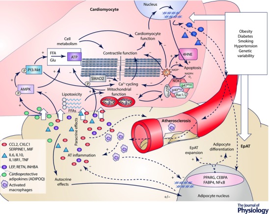
Figure 4. Communication between the cardiomyocytes and epicardial adipose tissue.

Epicardial adipose tissue (EpAT) and the cardiomyocyte transcriptomic profile are altered in the presence of cardiovascular risk factors or by genetic variability. Nevertheless, further to any systemic effects, a local cross‐talk takes place between cardiomyocytes and EpAT which determines aspects of myocardial biology, cardiac function and coronary atherosclerosis progression. Secreted adipokines (e.g. adiponectin or leptin) differentially affect AMP‐activated kinase (AMPK) and phosphoinositide 3‐kinase (PI3K)/Akt signalling in cardiomyocytes, which are centrally involved in cardiomyocyte metabolism and substrate utilization. Free fatty acid (FFA)‐related lipotoxicity results in mitochondrial dysfunction, impaired oxidative metabolism and increased oxidative stress. NADPH oxidase activity is also enhanced by mitochondrial dysfunction, and alterations in AMPK signalling induced by EpAT‐secreted adipokines. Increased phsopshorylation of SMAD2 (e.g. by activin A) and/or reduced PI3K/Akt signalling by pro‐inflammatory adipokines negatively affect Ca2+ cycling and cardiomyocyte contractility. Increased cardiomyocyte oxidative stress has also direct effects on redox‐sensitive proteins of the contractile apparatus and cell apoptosis. Cardiomyocyte stress due to impaired substrate utilization, contractile dysfunction and increased oxidative burden leads to respective changes in cardiomyocyte transcriptome. Products of increased myocardial oxidative stress, such as 4‐hydroxynonenal (4HNE, an end product of lipid oxidation) and possibly others among the cardiomyocyte secretome may signal back to EpAT and affect key aspects of its biology, such as the differentiation of adipocytes, adipose tissue expansion and its infiltration by inflammatory cells as well as the regulation of transcriptional factors and relevant gene expression profile, which is shifted towards a pro‐inflammatory phenotype. The concept of a bidirectional signalling between the heart and EpAT is represented with continuous (‘outside‐to‐inside’ signalling) and dashed arrows (‘inside‐to‐outside’ signalling) respectively. ADIPOQ, adiponectin; CEBPA, CCAAT/enhancer‐binding protein α; CXCL1, C‐X‐C motif chemokine ligand 1; FABP4, fatty acid binding protein‐4; IL, interleukin; CCL2, C‐C motif chemokine ligand; INHBA, activin A; LEP, leptin; MIF, macrophage migration inhibitory factor; NFκB, nuclear factor κB; PPARG, peroxisome proliferator activator receptor γ; RETN, resistin; SERPINE1, serpin family E member 1; SMAD2, mothers against decapentaplegic homolog 2; TNF, tumour necrosis factor α; list of adipokines is indicative. +, stimulate/induce; –, decrease/impair; short white arrows represent diffusion of adipokines.
