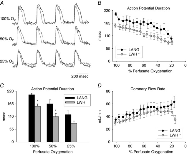
Figure 3. Changes in APD and coronary flow in LANG and LWHs during gradual perfusate deoxygenation when pacing at 330 ms CL.

A, representative optical APs for a LANG heart (black) and a LWH (grey) at 100%, 50% and 25% perfusate oxygenation. B, APD decreases with perfusate deoxygenation in both LWHs and LANG hearts. APDs during perfusate deoxygenation were lower in LWHs than LANG hearts. C, APD disparity between LWHs and LANG hearts at 100%, 50% and 25% perfusate oxygenation. APDs for LWHs were significantly less than LANG hearts at 100% and 50% perfusate oxygenation. D, CFR increases with perfusate deoxygenation in LWHs and LANG hearts. CFR during perfusate deoxygenation was lower in LWHs than LANG hearts. Values are the mean ± SE; LANG n = 7; LWH n = 7. *Significant difference between LANG and LWHs.
