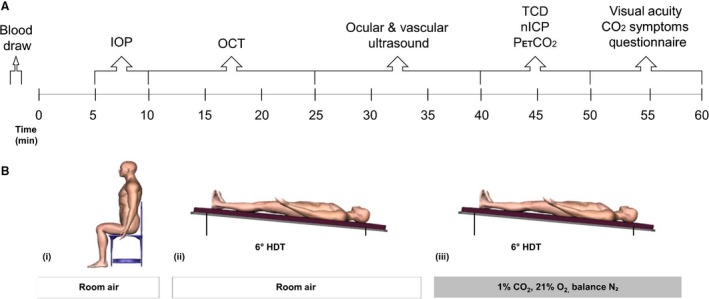
Figure 1.

Schematic of study protocol depicting (A) the timeline and order in which measurements were obtained during each condition and (B) the (i) Seated, (ii) HDT, and (iii) HDT + CO2 conditions. IOP, intraocular pressure; OCT, optical coherence tomography; TCD, transcranial Doppler ultrasound; nICP, noninvasive measure of intracranial pressure; PETCO 2, end‐tidal partial pressure of carbon dioxide.
