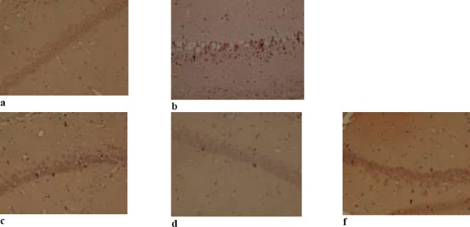
Figure 4.

Congo red staining to detect beta-amyloid plaques in the hippocampus in five group: (a) phosphate buffered saline (control group), (b) Aβ (50 ng/side), (c) Aβ (50 ng/side) + Iris (100 mg/kg), (d) Aβ (50 ng/side) + Iris (200 mg/kg), (f) Aβ (50 ng/side) + Iris (400 mg/kg).
