
Fig. 3. Characteristic structural features of muscle fibers are conserved after CLARITY treatment of EDL muscles while some modifications of fat cells and tendons are observed.
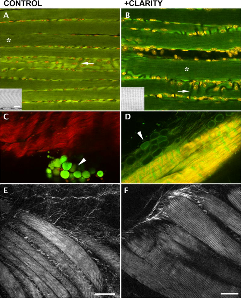
A–B: 2P fluorescence shows characteristic striated autofluorescence in both control and treated fibers. The insets reproduce the areas marked with an asterisk. Nuclei are positioned, as expected, along plasma membrane and along blood vessels that appear as dark channels (arrows); (C–D): SHG imaging (red) and 2-photon autofluorescence (green) of tendon and fat cells (arrowheads) show that the former keep their basic organization while fat cells are depleted and flattened by the treatment; E–F: SHG imaging of unstained EDL fiber bundles shows conservation of the striated pattern. Bars: A–E: 50 µm (see bar in E); F: 25 µm; insets 10 µm (bar in A).