
Figure 5. The age‐dependent decline and slowing of contraction is graded by frailty in voltage‐clamped ventricular myocytes.
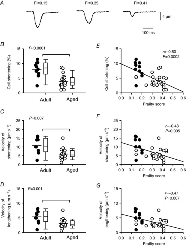
A, representative recordings of myocyte contractions recorded from mice with different FI scores. B, peak contractions normalized to cell size were smaller in aged animals compared to adults. C and D, velocities of shortening and lengthening were slower in the aged group compared to the adult group. E–G, scatterplots show that peak contractions, as well as the velocities of shortening and lengthening, were inversely proportional to FI score. Differences between age groups were assessed with a t test and correlations were assessed by linear regression (n = 10 adult and 24 aged myocytes). Filled symbols indicate adult mice and open symbols indicate aged mice.