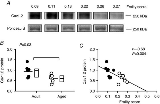
Figure 10. Frailty grades the age‐dependent decline in expression of Cav1.2 protein in the mouse heart.

A, representative Western blots for Cav1.2 protein expression in mice with varying FIs. Ponceau S staining was used as a loading control in all experiments (lower panels). B, mean expression of Cav1.2 decreased with age in the mouse heart. C, cardiac Cav1.2 expression was closely graded by frailty score in the mouse. Differences between age groups were assessed using a Mann–Whitney Rank Sum test and correlations were evaluated with linear regression analysis (n = 8 adult and 8 aged hearts). Filled symbols indicate adult mice and open symbols indicate aged mice.
