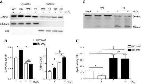
Figure 5.
GAPDH activated Ape1. WT and R3 SMCs were grown until confluence and treated with H2O2 (110 μM, 16 h). Cytosolic and nuclear fractions of WT and R3 cells were separated with NE-PER nuclear and cytoplasmic separation kit. A) Samples were immunoblotted with Ab for GAPDH, α-tubulin (cytosolic marker), and p53 (nuclear marker). B) Quantitative data show relative GAPDH amount in cytosolic and nuclear fraction (n = 3). *P < 0.05, ¶P < 0.01, §P < 0.005. C, D) Ape1 activity in nuclear fraction of WT and R3 cells. C) The 5′-endonuclease activity of Ape1 was quantified by measuring the incision of a 26-mer FAM-labeled oligonucleotide substrate that contained a single synthetic AP site. Ape1 endonuclease reaction mix contained 300 ng cell lysate and 1 μM FAM-labeled Ape1 substrate. Products were separated on 15% PAGE/Urea gels. D) Endonuclease activity was calculated as the relative amount of the 13-mer oligo product with the unreacted 26-mer substrate [product/(product + substrate)]. Data from a representative experiment are shown (n = 5). NS, not significant. *P < 0.05, §P < 0.005.

