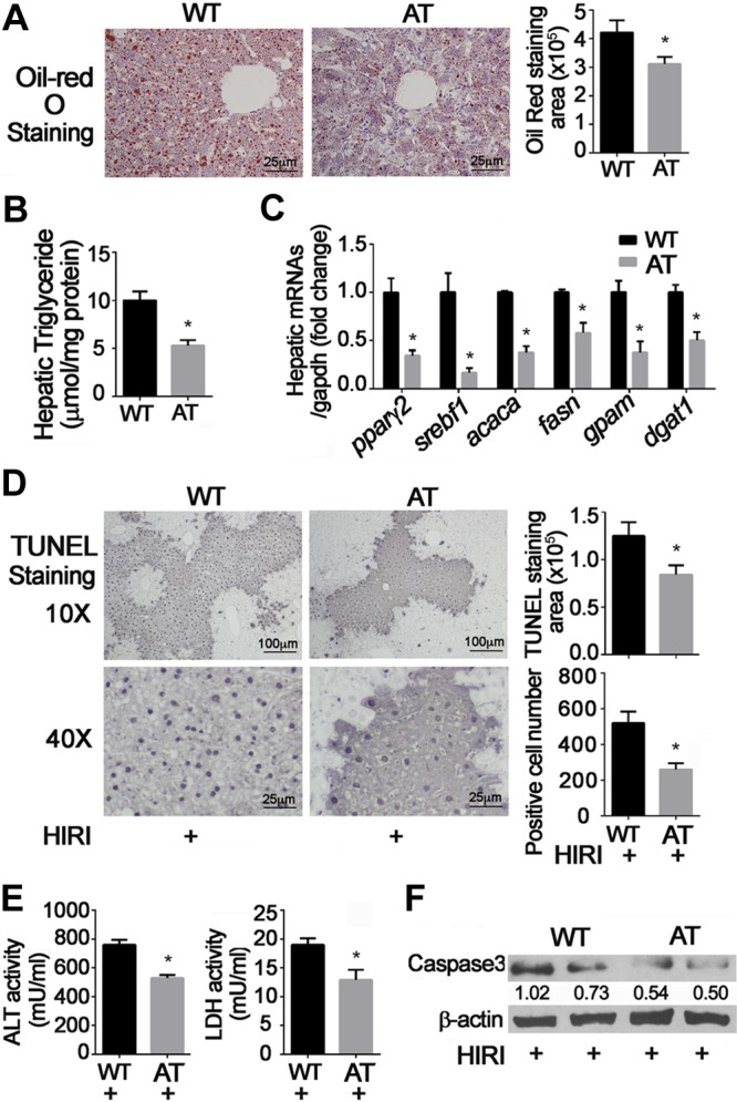
Figure 3.

Effects of mTOR activation and HFD on HIRI. AT mice were generated by cross-breeding Alb-Cre mice with TSC1floxp/floxp mice. Four-week-old animals were fed HFD for 12 wk, then challenged with HIRI. Results are expressed as means ± sem; n = 10 for each group. *P < 0.05 vs. wild-type mice; #P < 0.05 vs. AT mice (Student’s t test). A) Hepatic lipid content was measured by Oil Red O staining. B) Hepatic triglyceride content. C) mRNA levels of hepatic lipid metabolism–related genes were detected and normalized by glyceraldehyde phosphate dehydrogenase (GAPDH); D) TUNEL staining and number of positive hepatocytes shown at low (×10) and high (×40) magnification. E) Serum levels of ALT and LDH. F) Cleaved caspase 3 was detected by Western blot. β-Actin was used as internal control. Representative results from 3 independent experiments are shown.
