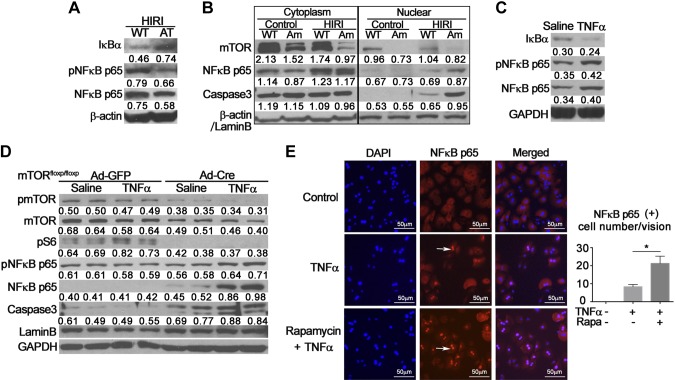
Figure 5.
Effects of hepatic mTOR signaling on nuclear translocation of NF-κB p65. A) Hepatic nuclear proteins from AT mice with HIRI were extracted. Western blot analysis was performed to detect alterations of IκBα and NF-κB p65. β-Actin was used as internal control. Representative data from 3 individual experiments are shown. B) Hepatic proteins from AM mice were extracted and separated into cytoplasmic and nuclear fractions. Western blot analysis was performed to validate knockdown of mTOR and nuclear translocation of NF-κB p65. β-Actin was used as internal control of cytoplasmic proteins. Lamin B was used as internal control of nuclear proteins. Western blot was repeated 3 times using distinct samples. C) Hepatocytes isolated from wild-type mice were treated with TNF-α (100 ng/ml) or saline control for 12 h. Western blot was performed to measure changes in IκBα and NF-κB p65. Glyceraldehyde phosphate dehydrogenase (GAPDH) was used as internal control. Experiment was repeated at least 4 times. D) Hepatocytes isolated from mTORfloxp/floxp mice were treated with Ad-GFP or Cre (5 × 10⁶ pfu/well) for 36 h, then exposed to TNF-α (100 ng/ml) for 12 h with saline as control vehicle. Western blot analysis was used to validate knockdown of mTOR and phosphorylation of NF-κB p65 and caspase 3. Lamin B and GAPDH were used as internal control. Experiment was repeated 3 times. E) Hepatocytes from wild-type mice were treated with TNF-α (100 ng/ml) with or without rapamycin (1 nM) for 12 h, and then probed for NF-κB p65 nuclear translocation by immunofluorescent staining. Positive nuclei indicated by arrows. NF-κB p65-positive nuclei were counted and expressed as means ± sem (n = 5). Experiment was repeated twice. P < 0.05 vs. TNF-α alone (Student’s t test).

