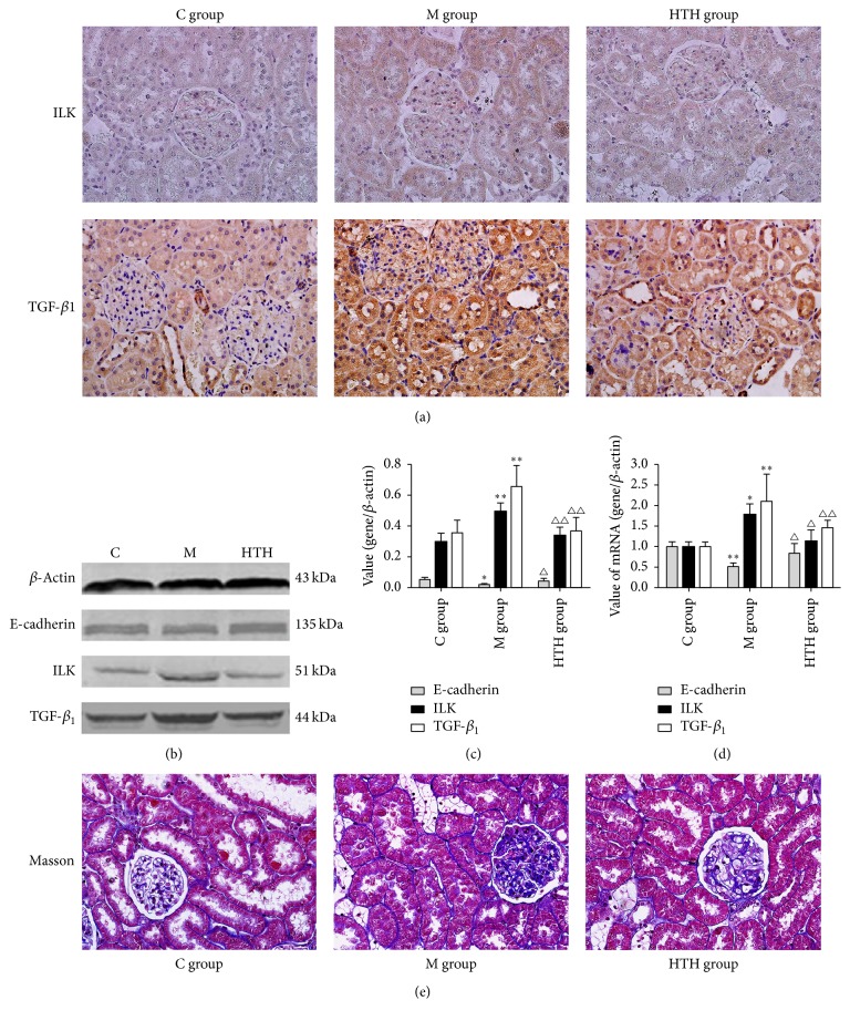
Figure 4.
Effects of HTH on E-cadherin, ILK, and TGF-β1 of rats. Immunohistochemical staining for ILK and TGF-β1 in the kidneys of rat of normal control rat group (C group), diabetic model rat group (M group), and Huayu Tongluo herbs treatment rat group (HTH group), exposed to DM and then administration of Chinese medicine of stasis removing and collaterals dredging herbal granule suspension intragastrically (a). Western blot detection and statistical analyses for renal E-cadherin, ILK, and TGF-β1 in kidneys of rat of three groups ((b), (c)). Quantification of E-cadherin, ILK, and TGF-β1 mRNA expression levels in different groups by Real-time PCR (d). Masson trichrome staining revealed extracellular matrix deposition in the kidneys of rat of three groups (stained in blue) (e). Values are expressed as means ± SD. Compared with C group, ∗P < 0.05, ∗∗P < 0.01; compared with M group, △P < 0.05, △△P < 0.01.

