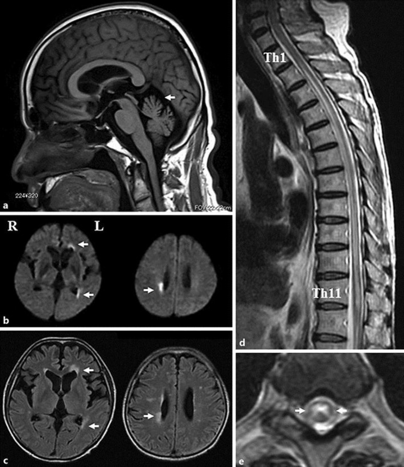
Fig. 1.
Sagittal T1-weighted MRI showing cerebellar atrophy (a, arrow). Axial diffusion-weighted (b) and fluid-attenuated inversion recovery (c) MRI showing high signals in the right posterior horn as well as the left anterior and posterior horns (arrows). Sagittal thoracic spine T2-weighted MRI showing high signal extending from Th1/2 to Th11 (d). Axial thoracic spine T2-weighted MRI showing high signal in the central part of the cord at the Th6/7 level (e, arrows).

