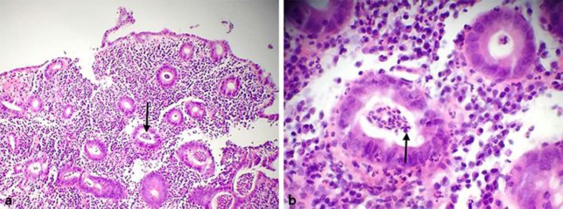
Fig. 1.
a Low-power view (10×) of January 2016 colonic mucosal biopsy of the patient showing diffuse active colitis with dense neutrophilic infiltrates producing crypt distortion, cryptitis, and crypt abscesses (arrow) consistent with the diagnosis of ulcerative colitis. b High-power view (40×) of the same colonic biopsy (January 2016) showing dense chronic inflammatory infiltrates of lymphocytes, plasma cells, and eosinophils (arrow). (Soriano and Soriano; histopathology by Antonio Alvarez Mendoza, MD.)

