
Figure 7. The impact of deficiency of CD4 and CD8 coreceptors on SEB-induced acute immunopathology in the lungs and livers of HLA-DR transgenic mice.
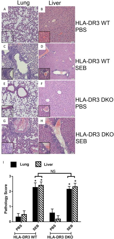
WT and DKO HLA-DR3 transgenic mice of either sex (8-10 weeks-old) were acutely challenged with PBS or SEB. Three days later, mice were euthanized, organs were collected, formalin fixed and paraffin embedded. H&E stained sections prepared from these tissue blocks were evaluated by light microscopy. Representative images are shown. Insets within each panel show higher magnifications. Panels A and B – Respective lung and liver sections from WT HLA-DR3 transgenic mice challenged with PBS. Panels C and D – Respective lung and liver sections from WT HLA-DR3 transgenic mice challenged with SEB. Panels E and F – Respective lung and liver sections from DKO HLA-DR3 transgenic mice challenged with PBS. Panels G and H – Respective lung and liver sections from DKO HLA-DR3 transgenic mice challenged with SEB. Panel I shows the histopathology scores which were determined as described in methods section. Each bar represents mean±SE values from 4-6 mice/group. * p<0.05 when compared to respective PBS-treated mice and NS – Not significant.