Fig. 2.

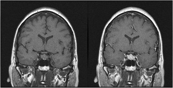
Pituitary MRI study. Left: T1 without contrast, Right: T1 with gadolinium contrast. Pituitary gland is symmetrically enlarged and enhances homogenously with gadolinium contrast

Pituitary MRI study. Left: T1 without contrast, Right: T1 with gadolinium contrast. Pituitary gland is symmetrically enlarged and enhances homogenously with gadolinium contrast