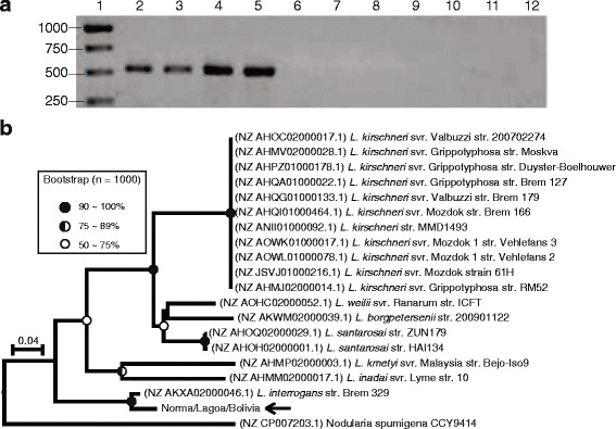
Fig. 3.

a PCR detection in Leptospira isolates and reference strains and (b) phylogenetic analysis of ORF36. The lanes in the gel (a) correspond to (1) molecular size marker, (2) svr. Hardjo (OMS), (3) isolate Norma, (4) isolate Lagoa, (5) isolate Bolivia, (6) svr. Lai, (7) svr. Copenhageni, (8) svr. Mini, (9) svr. Hebdomadis, (10) svr. Tarassovi, (11) svr. Sponselee and (12) negative control. The phylogenetic tree (b) was inferred using the neighbor-joining method (bootstrap: 1000, model: maximum composite likelihood) and rooted in the midpoint. Samples representing our clinical isolates are indicated with arrows. Circles at the nodes reflect the bootstrap support. An ancestral node without a circle indicates a bootstrap value below 50%
