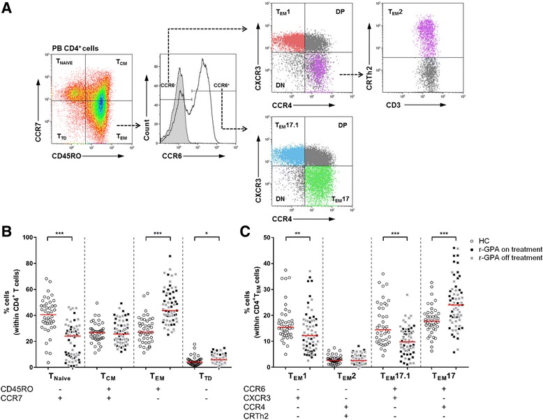
Fig. 1.

T cell subset distribution between HC and r-GPA patients. a Gating strategy of CD4+ TEM cell subsets using chemokine receptor expression patterns. The flow cytometry plots show sequential gating (dashed arrows) to identify different CD4+ TEM cell subsets within the CD4+TEM cell population. CD4+ T cell subsets from peripheral blood were identified based on the expression of CCR7 and CD45RO. Within CD4+TEM cells CCR6- and CCR6+ cells were identified based on the isotype (grey histogram with dashed line). Within CCR6- CD4+TEM cells expression of CXCR3 and CCR4 was analyzed to identify TEM1 cells (CD4+CD45RO+CCR7-CCR6-CXCR3+CCR4-). Expression of CRTh2 was used to identify lineage-committed TEM2 cells (CD4+CD45RO+CCR7-CCR6-CXCR3-CCR4+CRTh2+) derived from CCR6-CXCR3-CCR4+ CD4+TEM cells. The CXCR3 and CCR4 expression was also analyzed on CCR6+ CD4+TEM cells to identify TEM17 cells (CD4+CD45RO+CCR7-CCR6+CXCR3-CCR4+), and TEM17.1 cells (CD4+CD45RO+CCR7-CCR6+CXCR3+CCR4-). The unclassified populations within the CCR6- and CCR6+ populations were identified, that were double-negative (DN) or double-positive (DP) for CXCR3 and CCR4. Representative flow cytometry plots from one r-GPA patient. b Percentages of CD45RO-CCR7+ (T NAIVE), CD45RO+CCR7+ (T CM), CD45RO+CCR7- (T EM) and CD45RO-CCR7- (T TD) subpopulations within the CD4+ T cell population in peripheral blood of HCs (open circles; n = 42) and r-GPA patients (filled squares; n = 63). c The percentage of CD4+ TEM cell subsets in peripheral blood of HCs (open circles; n = 42) and r-GPA patients (filled squares; n = 63). Black squares indicate r-GPA patients on treatment (n = 29), grey squares indicate r-GPA patients off treatment (n = 34). Horizontal lines represent median percentages. * p < 0.05, ** p < 0.01, and *** p < 0.001. CCR C-C chemokine receptor, CD cluster of differentiation, CXCR3 CXC chemokine receptor 3, HC healthy control, r-GPA GPA patient in remission, T EM effector memory T cell
