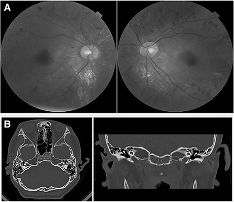
Fig. 2.

a Ophthalmoscopy photos of the patient showed the partial degeneration of the choroid in both eyes. b High-resolution CT imaging showed the cochlea deformity of the patient, including the short base turn of the cochlea, unclear division between the second turn and the apical turn of the cochlea and the absence of the modiolus
