
Figure 1.
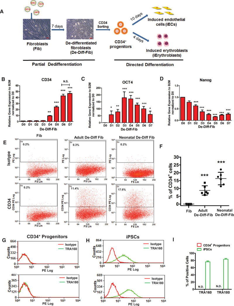
Generating CD34+progenitors from human dermal fibroblasts. A, Schematic of converting human dermal fibroblasts into induced endothelial cells (iECs) and induced Erythroblasts (iErythroblasts) by sorted CD34+ progenitors from dedifferentiated fibroblasts (De-Diff-Fib) at day 7. B through D, Quantitative polymerase chain reaction (PCR) analysis of the mesodermal marker CD34 (B), pluripotency markers OCT4 (C), and Nanog (D) during adult fibroblast dedifferentiation from days 0 to 7 (n=4). E, Representative flow-cytometry plots of the mesodermal marker CD34+ in fibroblasts, adult De-Diff-Fib, and neonatal De-Diff-Fib. F, Quantification and statistical analysis of CD34+ cells in fibroblasts (n=3), adult De-Diff-Fib (n=6), and neonatal De-Diff-Fib (n=6) from flow cytometry. G through I, Representative flow-cytometry overlay plots of the pluripotency markers: human TRA-160 and TRA-180 in CD34+ progenitors (G) and iPSCs (H) and quantification of TRA-160 or TRA-180-positive cells (I) in CD34+ progenitors and iPSCs. Data are presented as mean±SE, *P<0.05, **P<0.01, ***P<0.001, compared with day 0 in B, C, and D, compared with fibroblasts in F, using repeated measures 1-way analysis of variance in B, C, and D and using 1-way analysis in F. N.D. indicates not detected in I.