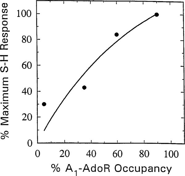
Fig. 10.

Correlation between SH interval prolongation and A1-AdoR occupancy by m-DITC-ADAC in guinea pig isolated, perfused hearts. Hearts paced at a constant atrial cycle length of 300 msec were perfused with Krebs-Henseleit solution containing 5 μM m-DITC-ADAC for 5, 10, 15, and 30 min. At the end of each perfusion period, the tissue was perfused with 5 μM CPT, and the irreversible component of SH interval prolongation was recorded. After washout for 1 hr, the ventricles were harvested, and membranes were prepared for determination of the number (Bmax) and affinity (Kd) of A1-AdoRs by measuring specific binding of [3H]CPX. The irreversible component of SH interval prolongation caused by m-DITC-ADAC is plotted as a function of percent of total A1-AdoRs occupied [Bmax (fmol/mg of protein of untreated control)/Bmax of m-DITC-ADAC-treated hearts × 100). The Bmax and Kd values of four untreated hearts were 33 ± 4 fmol/mg of protein and 3.2 ± 0.2 nM, respectively. There was no significant difference between the Kd values of [3H]CPX binding to membranes of untreated and m-DITC-ADAC-treated hearts.
