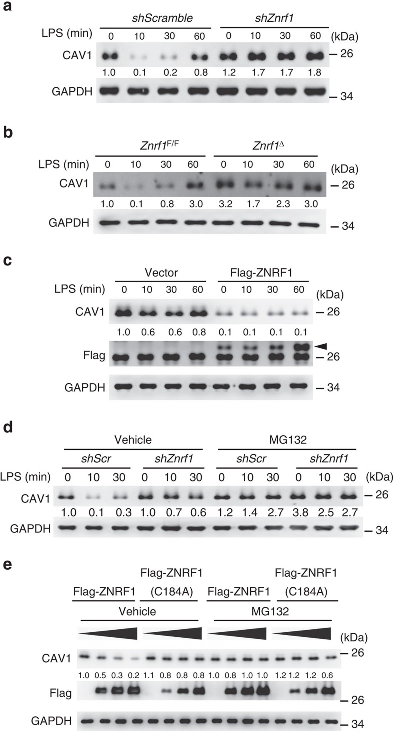
Figure 3. ZNRF1 regulates CAV1 protein stability.
(a) Immunoblot analysis of CAV1 and GAPDH in lysates of control (scrambled shRNA) and ZNRF1-shRNA-expressing RAW264.7 cells stimulated with LPS (100 ng ml−1) for the indicated times. (b) Immunoblot analysis of CAV1 and GAPDH in lysates of Znrf1F/F and Znrf1Δ cells treated with LPS (100 ng ml−1) for the indicated times. (c) Immunoblot analysis of the indicated proteins in lysates of control (vector) and Flag-tagged ZNRF1-expressing RAW264.7 macrophages stimulated with LPS (100 ng ml−1). The arrow indicates Flag-ZNRF1. (d) Immunoblot analysis of CAV1 protein in lysates of scrambled control and Znrf1-knock-down RAW264.7 cells stimulated with 100 ng ml−1 LPS with or without pretreatment with MG132 (10 μM). (e) HEK293T cells were transfected with increasing amounts of Flag-tagged wild-type ZNRF1 or ZNRF1(C184A); 24 h after transfection, the cells were treated with MG132 (10 μM) for at least 6 h, and cell lysates were analysed by immunoblotting with the indicated antibodies. The intensities of the bands are expressed as fold increases compared to those of untreated control cells after normalization to GAPDH expression. The data are representative of three independent experiments performed in triplicate.

