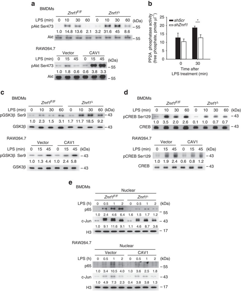
Figure 6. Akt–GSK3β–CREB signalling is altered in ZNRF1-deficient and CAV1-expressing macrophages in response to LPS.
(a,c,d) Immunoblot analysis of the phosphorylation of Akt (Ser473), GSK3β (Ser9) and CREB (Ser129) as well as total Akt, GSK3β, and CREB in lysates of Znrf1F/F and Znrf1Δ BMDMs and vector- and CAV1-expressing RAW264.7 cells stimulated with LPS (100 ng ml−1) for the indicated times. (b) RAW264.7 macrophages infected with lentiviruses expressing shScr or shZnrf1 were treated with LPS (100 ng ml−1) for 30 min, and cell lysates were prepared and PP2A catalytic subunit was immunoprecipitated followed by PP2A phosphatase activity analysis. (e) Immunoblot analysis of NF-κB p65 and c-Jun in nuclear extracts isolated from BMDMs and RAW264.7 cells described above. Histone H3 served as a marker for the nuclear fraction. The intensities of the bands are shown as fold increases compared to those of untreated control cells after normalization to their unphosphorylated forms or H3 expression. *P<0.001 (Student's t-test). The data are representative of three independent experiments performed in triplicate (error bars, s.d.).

