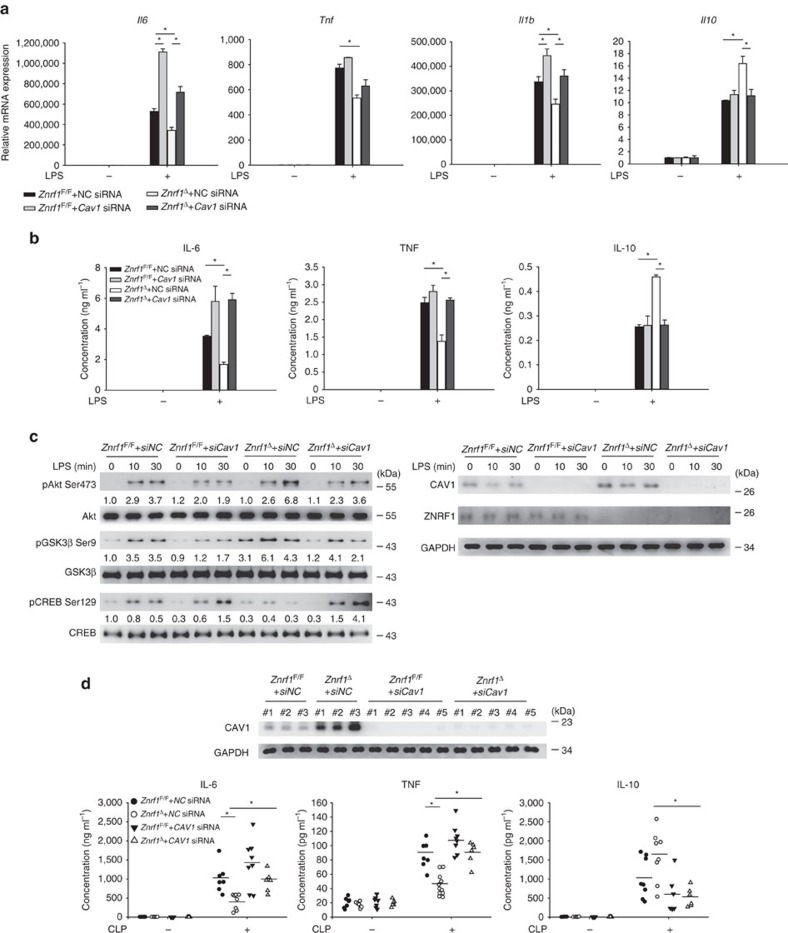
Figure 7. ZNRF1-mediated TLR4- and CLP-triggered inflammatory responses depend on CAV1.
(a–c) Znrf1F/F or Znrf1Δ BMDMs electroporated with either control (NC) or Cav1 siRNA were treated with LPS (100 ng ml−1). (a) The expression of the indicated mRNAs in BMDMs after stimulation with LPS for 4 h was analysed by RT–qPCR. (b) The production of cytokines in culture supernatants of BMDMs after treatment with LPS for 8 h was determined by ELISA. (c) Phosphorylation of Akt, GSK3β, and CREB as well as the indicated proteins in cell lysates were analysed by immunoblotting with the indicated antibodies. The intensities of the bands are shown as fold increases compared to those of untreated control cells after normalization to their unphosphorylated forms. The data are representative of three independent experiments performed in triplicate (error bars, s.d.). (d) Znrf1F/F or Znrf1Δ mice were injected intravenously with either control (NC) or Cav1 siRNA. After 36 h, mice were subjected to CLP, and blood was collected 8 h post CLP. The indicated cytokines in sera were determined by ELISA (bottom), and CAV1 protein level in peripheral blood leukocytes were analysed by immunoblotting (top). *P<0.05 (Student's t-test).

