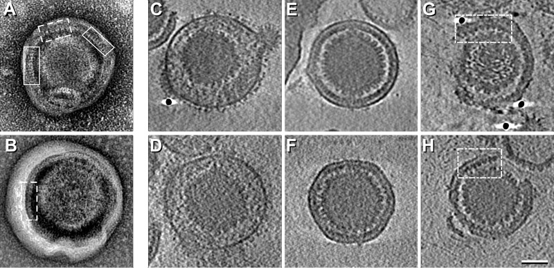
FIG 1 .
Electron micrographs and cryo-ET slices of PEVs. (A and B) Negatively stained micrographs showing “picket fence” motifs in the NEC layer. Tracts with the 6-nm repeat are boxed with continuous lines and tracts with the 10.5-nm repeat with dashed lines. (C to H) Examples of near-central slices through the respective density maps. (C and D) Mature virions. (E and F) Intact PEVs; (G and H) Broken PEVs. Tracts showing the 12-nm repeat are boxed. Scale bar = 50 nm.

