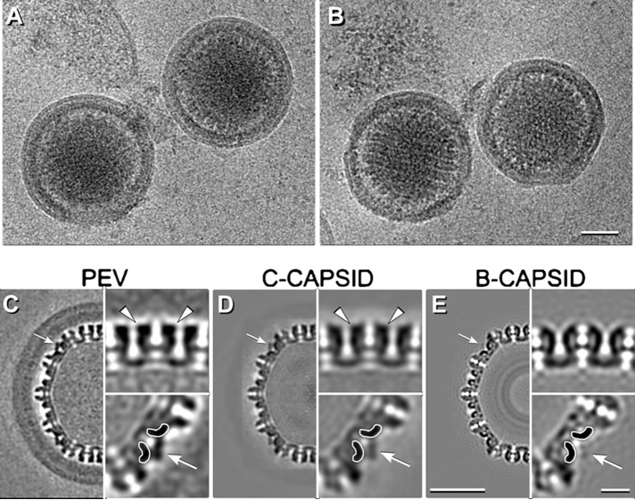
FIG 3 .
Cryo-EM reconstruction of PEVs. (A and B) Cryo-micrographs of intact PEVs. (C to E) Central sections of reconstructions of a PEV (C), a C-capsid (EMDB code 1354) (D), and an in vitro-matured B-capsid (27) (E). White arrows in panels C to E point to CCSC sites that are occupied in panels C and D but not in panel E. Symmetry-related regions are shown in magnified insets at bottom right in panels C to E. The triplexes to which the CCSC binds are delineated with white contours. The outcrops of density on hexamers indicative of pUL35 are marked with white arrowheads in panels C and D. Scale bars: panels B and D, 50 nm; inset in panel E, 10 nm.

