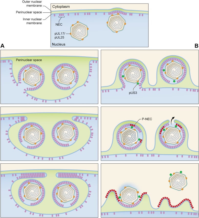
FIG 7 .
Pathways for nuclear egress in UL3-null and wild-type infections. In both cases, C-capsids start to bud at NEC-rich regions of the INM (top panel). (A) US3-null pathway leading to the formation of a nuclear sac. (B) Wild-type pathway. The top image in panel A shows one particle budded off with a “fontanelle” gap in its NEC layer and one particle at an advanced stage of budding. In the middle image, both PEVs have budded off and face-to-face interactions between apposing NEC layers are beginning to close off the sac. In the bottom image, this process has proceeded almost to completion. (B) In the wild-type pathway, we envisage that proteins entering the interstitial space between the NEC layer and capsid include the pUS3 kinase (green balls) and assume that there is no major structural difference between these PEVs and US3-null PEVs. Some pUS3 may be accommodated in the fontanelle gap; in any case, pUS3 should be sufficiently mobile to be able to access and phosphorylate the NEC layer (magenta-to-red switch), which would consequently disengage from the capsid. The fusion event that releases the capsid into the cytoplasm (middle image) would be facilitated by having the fontanelle membrane, which is presumably more pliable than the NEC-backed membrane, apposed to the ONM (black arrow). Finally (bottom image), the NEC proteins (P-NEC) are no longer bound to the capsid, which goes on to later stages of maturation, including acquisition of tegument proteins and secondary envelopment (not shown).

