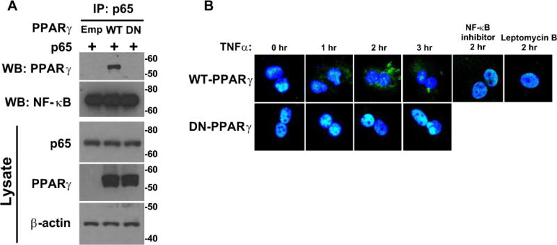
Figure 5. PPARγ Association with p65.

A) HEK293T cells transfected with p65, WT-PPARγ or DN-PPARγ were treated with a proteasome inhibitor, MG132 (5 μM, 12 hr). Proteins were immunoprecipitated with p65 antibody and immunoprecipitated proteins were Western blotted for the indicated protein. The top 2 blots represent immunoprecipitation with p65 and Western blot with the indicated antibody. The bottom 3 blots represent Western blots for the indicated protein from cell lysates. Size markers transferred from the blots are shown. B) PPARγ immunostaining (green) of WT-PPARγ or DN-PPARγ expressing SMC treated with TNFα for the indicated times. Where indicated, cells were treated with an NF-κB inhibitor (50 μMr) or leptomycin B (5 nM) for 1 hour prior to TNFα.
