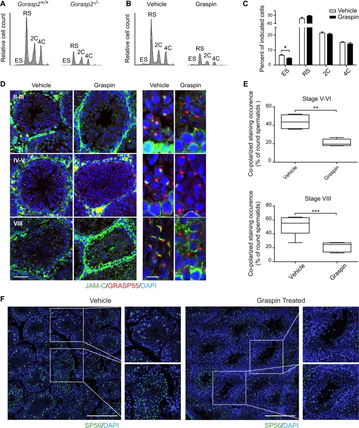
Fig 7. Chemical inhibition of GRASP55 affects spermatid differentiation in vivo.
(A-B) Flow cytometry profiles of DAPI-stained germ cells isolated from testes of Gorasp2+/+ and Gorasp2-/- 35-days old mice (A) and 27-days old mice treated with vehicle or Graspin for two weeks (B). ES: elongated spermatids, RS: round spermatids, 2C: spermatogonia, 4C: primary spermatocytes. (C) Quantification of germ cell numbers of 27-days old mice treated with Graspin for two weeks and analyzed two days after the last Graspin injection. Vehicle, n = 10; Graspin, n = 10. Student’s unpaired t-test; *: P<0.05. (D) Confocal images of JAM-C, GRASP55 and DAPI staining of seminiferous tubule sections of 43-days old mice treated for 16 days with vehicle or Graspin. Differentiation stages of seminiferous tubules are indicated. High magnification pictures on the left panel highlight the loss of co-polarized localization of GRASP55 and JAM-C in developing round spermatids. Scale bars: left panels: 50 μm; right panels: 10 μm. (E) Quantification of co-polarized JAM-C and GRASP55 staining occurrence in control and treated mice expressed as the percentage of round spermatids showing a close apposition between GRASP55 and JAM-C stainings. Results obtained upon quantification of co-polarized staining occurrences in seminiferous tubules at stage V-VI (n = 415 counted events, n = 4 mice, 2 tissue sections/mice) and VIII (n = 498 counted events, n = 6 mice, 2 tissue sections/mice) are shown. Student’s unpaired t-test; **: P<0.01, ***: P<0.001. (F) Confocal images of SP56 and DAPI staining of seminiferous tubule sections of 43-days old mice treated for 16 days with vehicle or Graspin as indicated. High magnification pictures on the right panels highlight the loss of SP56 staining associated with elongated spermatids. Scale bars: 200 μm.

